Showing posts with label echocardiogram. Show all posts

Oct 20, 2023

 Understanding Echocardiography: Peering into the Heart's Inner Workings


Introduction:
Echocardiography, often referred to as an "echo" in the medical field, is a non-invasive diagnostic test that uses high-frequency sound waves (ultrasound) to produce real-time images of the heart. It has revolutionized the way healthcare professionals assess the structure and function of the heart, making it an essential tool in the field of cardiology. This article will delve into the world of echocardiography, exploring its uses, techniques, and the crucial role it plays in diagnosing and managing cardiovascular conditions.

The Basics of Echocardiography:
Echocardiography is a painless and safe imaging procedure that offers a dynamic view of the heart. It operates on the same principle as the ultrasound used in prenatal imaging but is tailored to assess the heart. The primary types of echocardiography include:

  1. Transthoracic Echocardiography (TTE): This is the most common form of echocardiography. It involves applying a gel on the patient's chest and using a transducer to capture images through the chest wall. TTE provides an overall view of the heart's structure and function.

  2. Transesophageal Echocardiography (TEE): TEE is a more invasive procedure that involves inserting a specialized transducer into the esophagus to obtain clearer images, particularly for patients with challenging anatomy or conditions.

Applications of Echocardiography:
Echocardiography serves various purposes in the field of cardiology:

  1. Diagnosis: It helps diagnose heart conditions such as valvular diseases, congenital heart defects, cardiomyopathies, and more.

  2. Assessment of Heart Function: Echocardiography provides crucial information about the heart's size, shape, and pumping ability.

  3. Guidance for Procedures: Cardiologists use echocardiography to guide procedures such as cardiac catheterizations and heart surgeries.

  4. Monitoring: It's instrumental in monitoring the progression of heart diseases and the effectiveness of treatments.

Doppler Echocardiography:
Doppler echocardiography is an extension of standard echocardiography that assesses blood flow within the heart and blood vessels. It measures the speed and direction of blood flow, helping identify issues like regurgitation or stenosis of heart valves.

Echocardiography in Pediatric Care:
Echocardiography isn't limited to adults; it's a valuable tool in pediatric cardiology. It aids in diagnosing congenital heart defects in infants and children, allowing for early intervention and treatment.

Advancements in Echocardiography:
Echocardiography has seen significant technological advancements. Today, 3D echocardiography and strain imaging provide even more detailed and accurate information. These technologies enable healthcare providers to identify heart problems at an earlier stage and offer more personalized treatments.

Conclusion:
Echocardiography is a cornerstone of modern cardiology. Its non-invasive nature, coupled with its ability to provide comprehensive insights into the heart's structure and function, makes it an invaluable diagnostic tool. Whether diagnosing heart conditions, guiding interventions, or monitoring treatment outcomes, echocardiography plays a pivotal role in improving the health and well-being of countless individuals with cardiovascular issues.

As technology continues to advance, echocardiography remains at the forefront of cardiac care, contributing to better patient outcomes and the ongoing fight against heart disease.


Oct 19, 2023

Exploring the Wonders of An echocardiogram uses sound waves to create pictures of the heart. This common test can show blood flow through the heart and heart valves: Seeing the Heart in Action

 Exploring the Wonders of Echocardiography:


Seeing the Heart in Action


Exploring the Wonders of Echocardiography: Seeing the Heart in Action

Introduction

Echocardiography, often referred to as an "echo," is a powerful diagnostic tool used in the field of cardiology. It employs the principles of ultrasound technology to create detailed, real-time images of the heart, providing invaluable information about its structure, function, and overall health. This non-invasive, safe, and painless imaging technique has become a cornerstone in the diagnosis and management of heart conditions. In this article, we will delve into the fascinating world of echocardiography, exploring its various types, applications, and the pivotal role it plays in modern medicine.

Understanding Echocardiography

Echocardiography is a medical imaging technique that uses high-frequency sound waves to produce images of the heart. The basic process can be summarized in a few key steps:

  1. Sound Wave Emission: A transducer emits high-frequency sound waves (ultrasound) that bounce off the heart's structures.

  2. Echo Reception: These sound waves echo back when they encounter various cardiac tissues, including the walls, valves, and chambers of the heart.

  3. Image Generation: A computer processes the returning echoes, translating them into detailed images displayed on a monitor.

Types of Echocardiography

  1. Transthoracic Echocardiography (TTE):

    • This is the most common type of echocardiography. It is non-invasive and involves placing the transducer on the chest's surface to obtain images of the heart through the chest wall.
  2. Transesophageal Echocardiography (TEE):

    • TEE provides even more detailed images by inserting a specialized transducer into the esophagus. It is especially useful for visualizing the posterior structures of the heart.
  3. Doppler Echocardiography:

    • This technique measures blood flow within the heart and blood vessels. It helps in assessing the speed and direction of blood flow, which is crucial for diagnosing conditions like heart valve abnormalities.

Applications of Echocardiography

Echocardiography plays a pivotal role in the diagnosis, assessment, and management of various heart conditions:

  1. Cardiac Anatomy and Function Evaluation:

    • It helps visualize the heart's chambers, walls, valves, and blood flow, providing crucial information about the heart's structure and how it is functioning.
  2. Diagnosis of Heart Diseases:

    • Echocardiography is used to diagnose conditions such as heart valve disorders, congenital heart defects, heart muscle diseases, and pericardial diseases.
  3. Monitoring Heart Health:

    • It allows physicians to monitor the progression of heart conditions and assess treatment effectiveness.
  4. Guidance for Surgical Procedures:

    • Surgeons use echocardiography to guide them during heart surgeries and interventions.
  5. Assessment of Cardiac Function:

    • Echocardiography can help determine ejection fraction, a crucial parameter for understanding the heart's pumping ability.

Advancements in Echocardiography

Echocardiography has advanced significantly over the years. Some notable developments include:

  1. 3D and 4D Echocardiography: These technologies provide even more comprehensive images of the heart, aiding in complex structural assessments and surgical planning.

  2. Contrast-Enhanced Echocardiography: Contrast agents are used to improve visualization and assess blood flow in the heart.

  3. Speckle Tracking Echocardiography: This technique measures subtle changes in the heart's muscle motion, enabling early detection of heart problems.

Conclusion

Echocardiography has become an indispensable tool in the field of cardiology. Its ability to offer real-time, high-quality images of the heart, without invasive procedures, has transformed the way heart conditions are diagnosed and managed. As technology continues to advance, the potential for earlier and more accurate diagnoses and more precise treatment plans is on the horizon, ensuring that echocardiography will continue to be at the forefront of cardiovascular healthcare for years to come.

Oct 31, 2015

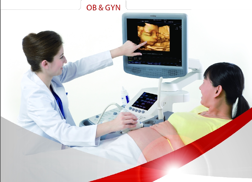
ultrasound system | DC-7 | Echo | 4D | mindray

DC-7    


Premium imaging Premium imaging platform technologies
Real-time 4D imaging
Power and Directional Color Doppler
Tissue Doppler imaging
Tissue harmonic imaging
IMT: intima-media thickness
Free Xros M: anatomical M mode
Steered scanning B, color/power, PW/CW independent
Trapezoid imaging
iScape: panoramic imaging
iBeam™ : spatial compounding imaging

Frequency compounding imaging

iClear™ : adaptive speckle suppression imaging
iTouch: one-key optimization for B, PW/CW  images

User productivity features:

High resolution 17 " LCD display on omni-directional  articulated  arm
Touch screen control panel
4 active transducer sockets
Home base user interface
Ergonomic and lightweight transducers
iTouch: one-key optimization for B, PW/CW  images
Multifunctional control
Best Pan and Spot combination ZOOM to visualize small anatomic structures
User programmable pre-sets for clinical specialties, patient types
User programmable measurement, calculation and operator preferences

Streamlined workflow to maximize efficiency

Fast system response: Four active probe  ports, iStation. DICOM3.0 (Storage, Print,DIR, Worklist, MPPS, HIS, RIS)

Special applications:

3D/4D imaging
IMT: automatic calculation and analysis of carotid intima-media thickness