Oct 31, 2015

ultrasound system | DC-N2 Color Doppler Imaging | MINDRAY

DC-N2


Mindray’s DC-N2 offers a unique combination of relevant technology, quality and affordability.
By integrating new platform and image processing algorithms,
the DC-N2 can be used by all across multiple clinical settings with the following new features:
DC-N2


  • Features :
    • 15-inch high resolution color LCD monitor
    • B-Mode
    • THI and PSH
    • M-Mode
    • Color Doppler Imaging
    • Integrated 320G hard drive
    • Power Doppler Imaging and Directional (PDI )
    • Pulsed Wave Doppler
    • HPRF (High Pulse Repeat Frequency)
    • iClearTM (Speckle Suppression Imaging)
    • iBeamTM (Spatial Compounding Imaging)
    • iTouchTM (Auto Optimization)
    • Zoom/iZoomTM  (Full Screen Zoom)
    • FCI (Frequency Compounding Imaging)
    • Phase Shift Harmonic imaging for all probes
    • iStationTM
    • iVisionTM
    • 2 active transducer connectors
    • DVD-RW Driver
    • 3 USB ports
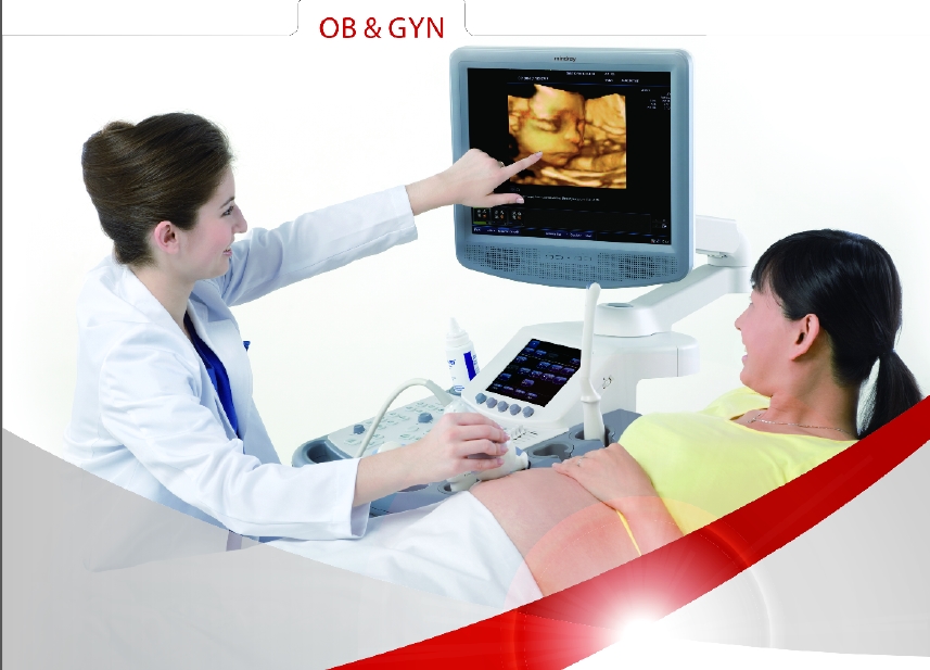
    • Shared Service Application Package (Abdominal, Obstetrical, Gynecological,  Cardiac, Small Parts, Urological, Vascular, Pediatric Packages)
    • Auto Doppler Calculation
    • iStorage (Direct Network Storage)
    Tutorial function:
    • iScanHelper
    • iClear : Speckle reduction imaging
    • iBeam: Spatial compounding imaging
    Main functions:
    √ iScanHelper: Mindray unique self-learning software
    √ Smart 3D: Freehand 3D imaging
    √ iScape: Real-time panoramic imaging
    √ Auto IMT: Automatically detect and calculate intima-media thickness
    √ iTouch: One-button image optimization
    √ iZoom Automatically expand the image to full screen
    √ iStorage : Direct data transfer to PC
Gallery
    

ultrasound system | DC-7 | Echo | 4D | mindray

DC-7    


Premium imaging Premium imaging platform technologies
Real-time 4D imaging
Power and Directional Color Doppler
Tissue Doppler imaging
Tissue harmonic imaging
IMT: intima-media thickness
Free Xros M: anatomical M mode
Steered scanning B, color/power, PW/CW independent
Trapezoid imaging
iScape: panoramic imaging
iBeam™ : spatial compounding imaging

Frequency compounding imaging

iClear™ : adaptive speckle suppression imaging
iTouch: one-key optimization for B, PW/CW  images

User productivity features:

High resolution 17 " LCD display on omni-directional  articulated  arm
Touch screen control panel
4 active transducer sockets
Home base user interface
Ergonomic and lightweight transducers
iTouch: one-key optimization for B, PW/CW  images
Multifunctional control
Best Pan and Spot combination ZOOM to visualize small anatomic structures
User programmable pre-sets for clinical specialties, patient types
User programmable measurement, calculation and operator preferences

Streamlined workflow to maximize efficiency

Fast system response: Four active probe  ports, iStation. DICOM3.0 (Storage, Print,DIR, Worklist, MPPS, HIS, RIS)

Special applications:

3D/4D imaging
IMT: automatic calculation and analysis of carotid intima-media thickness


ultrasound system | DC-70 | ilive = 5 D | Echo | Mindray


DC-70


Good image quality is prime for a promising ultrasound. DC-70 offers that and a lot more. Its unique touch gestures let you do more than you ever imagined, providing you with extremely easy and fast exams, enabling you to put patient care first. The clarity, as a result of high-end inherent technologies, would help you achieve easy fetal biometry analysis, 4D imaging, and GYN abnormalities detection with best in class image performance. The quality exams and unprecedentedly optimized user experience have already built up DC-70 as the true workhorse to cope with various challenges of your daily OB/GYN practice.


Ergonomics

• 19 inch LED monitor with articulated arm
• 10.4 inch gesture sensitive touch screen with adjustable angle
• Flexible control panel
• Gel warmer

Workflow

Gesture Sensitive Touch Screen The smart 10.4 inch motion sensitive touch screen lets you do more than you ever imagined. Be it scrolling through images, shifting the image to and from the monitor, zoom in/out, measure or starting specific user-defined functions with a defined hand motion, the DC-70 allows to get more done and much faster
.
Smart OB™
Auto measurement of fetal parameters: trace and calculate the BPD, OFD, HC, AC and FL on a single click
Smart-NT
Auto-trace tube cavity edge with measurement result
Smart-V
Automated trace measurements including accurate 3D VOI sketching and 3D volume size measurement
FreeView™
Improves patient’s comfort by automatically changing the angle of scanning plane on the Endo-cavity volume probe DE10-3E from -45° to + 45°
Raw Data
Enables optimum flexibility for post processing of the stored images including parameter adjustments, adding comments and measurements, allowing maximum productivity during scanning
iZoom™
Gain instant full screen view on the click of a single key
MedTouch
Size and physical distance now is no longer an obstacle. With MedTouch, a one-stop solution provides you with a smarter way to control the ultrasound device, access patient data and inbuilt tutorial software via your android operated smart device.
MedSight
DC-70 lets you transfer clinical images and cine to your IOS or android powered smart device via an interactive app. It could be for a to-be-mother wanting to share the images of the fetus with her family or friends, it could be a training session or a discussion with your peers on a rare case, with MedSight you can now take the clinical examinations with you wherever needed.


Performance

iLive
Integrating traditional ray casting algorithm with a new virtual lighting mode,iLive generates exciting real visual effects such as interactive shadowing, skin scattering and human skin-like images.
Page™
A CT-like view functionality displaying volume data in multiple parallel 2D images to effectively interpret anatomical structures
Curved MPR™
Providing complete visual examination for organs and structures of different shapes by 3D volume data view in linear and curved planes
SCV™
Slice Contrast View helps deliver higher contrast resolution and reduced noise, enabling higher-quality anatomical assessment with more information
Niche/3 Slice
Get a 3D sectional view of the A, B and C planes to better understand the internal structure 
Color STIC
Spatio-Temporal Image Correlation permits advanced fetal heart diagnosis with color flow in real time 4D
Color 3D
Analyze branched vessels effectively with 3D color Doppler

Gallery :


Oct 30, 2015

The latest ultrasound and X-ray equipment Cairo Medical Services Company

The latest ultrasound and X-ray equipment Cairo Medical Services Company and the Agent of Mindray Egypt
Cash and installment